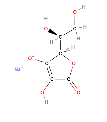
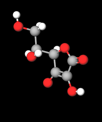
Best studies on sodium ascorbate
BACKGROUND:Vitamin C (L-ascorbic acid), a known enhancer of collagen deposition, has also been identified as an inhibitor of elastogenesis. OBJECTIVE: Present studies explored whether and how the L-ascorbic acid derivative (+) sodium L-ascorbate (SA) would affect production of collagen and elastic fibers in cultures of fibroblasts derived from normal human skin and dermal fat, as well as in explants of normal human skin, stretch-marked skin and keloids. METHODS: Effects of SA on the extracellular matrix production were assessed quantitatively by PCR analyses, western blots, biochemical assay of insoluble elastin and by immuno-histochemistry. We also evaluated effects of SA on production of the reactive oxygen species (ROS) and phosphorylation of IGF-I and insulin receptors. RESULTS: SA, applied in 50-200 μM concentrations, stimulates production of both collagen and elastic fibers in all tested cultures. Moreover, combination of SA with a proline hydroxylase inhibitor induces a beneficial remodelling in explants of dermal scars, resulting in the inhibition of collagen deposition and induction of new elastogenesis. Importantly, we revealed that SA stimulates elastogenesis only after intracellular influx of non-oxidized ascorbate anions (facilitated by the sodium-dependent ascorbate transporter), that causes reduction of intracellular ROS, activation of c-Src tyrosine kinase and the enhancement of IGF-1-induced phosphorylation of the IGF-1 receptor that ultimately triggers elastogenic signalling pathway. CONCLUSION:
Our results endorse the use of this potent stimulator of collagen and elastin production in the treatment of wrinkled and stretch-marked skin. They also encourage inclusion of SA into therapeutic combinations with collagenogenesis inhibitors to prevent formation of dermal scars and keloids (1).
There is mounting evidence demonstrating causative links between hyperglycemia, oxidative stress, and insulin resistance, the core pathophysiological features of type 2 diabetes mellitus. Using a combinational approach, we synthesized a vanadium-antioxidant (i.e., l-ascorbic acid) complex and examined its effect on insulin resistance and oxidative stress. This study was designed to examine whether vanadyl(IV)-ascorbate complex (VOAsc) would reduce oxidative stress, hyperglycemia, and insulin resistance in high-fat high-sucrose diet (HFSD)-induced type 2 diabetes in mice. Male C57BL/6J mice were fed a HFSD for 12 weeks to induce insulin resistance, rendering them diabetic. Diabetic mice were treated with rosiglitazone, sodium l-ascorbate, or VOAsc. At the end of treatment, fasting blood glucose, fasting serum insulin, homeostasis model assessment-insulin resistance index, and serum adipocytokine levels were measured. Serum levels of nitric oxide (NO) parameters were also determined. The liver was isolated and used for determination of malondialdehyde, reduced glutathione, and catalase levels, and superoxide dismutase and glutathione peroxidase activities. VOAsc groups exhibited significant reductions in serum adipocytokine and NO levels, and oxidative stress parameters compared to the corresponding values in the untreated diabetic mice. The results indicated that VOAsc is non-toxic. In conclusion, we identified VOAsc as a potentially effective adjunct therapy for the management of type 2 diabetes (2).
A comparative study on the antioxidant effect of rosemary extract (RE) and sodium ascorbate (SA) on lipid and colour oxidation of liver pâté made of lard and pork liver was done. During the 48 hour experimental time all the pâtés were wrapped in a foil and stored in cold room of 3.5°C under light of 1000 lux. Colour stability was monitored by instrumental colour measurement (CIE L*a*b* colour space) whereas lipid stability was measured by the determination of the 2-thiobarbituric acid reactive substances (TBARS). In the present study RE doses range (0,125, 250, 375 and 500 ppm) showed no significant (p>0.05) and linear effect on colour stability. However thez RE revealed a significant effect (p<0.05) against lipid oxidation and linearly reduces the TBARS number. The added SA doses (0, 250, 500, 750 and 1000 ppm) revealed significant (p<0.05) and linear effect in reducing discoloration. However the studied SA dose ranges showed no significant (p>0.05) effect on TBARS number. In this study RE was showed better performance against lipid oxidation and SA was potent against discoloration. The effect of the added spices used in manufacturing of the studied product showed no significant (p>0.05) effect against lipid and color oxidation. However the added spices revealed possible antagonistic and synergetic relationship with the studied the antioxidant systems (RE & SA) (3).
BACKGROUND: Neuroblastoma (NB) is an extra-cranial solid tumour of childhood. In spite of the good clinical response to first-line therapy, complete eradication of NB cells is rarely achieved. Thus, new therapeutic strategies are needed to eradicate surviving NB cells and prevent relapse. Sodium ascorbate has been recently reported to induce apoptosis of B16 melanoma cells through down-regulation of the transferrin receptor, CD71. Since NB and melanoma share the same embryologic neuroectodermal origin, we used different human NB cell lines to assess whether the same findings occurred. RESULTS: We could observe dose- and time-dependent induction of apoptosis in all NB cell lines. Sodium ascorbate decreased the expression of CD71 and caused cell death within 24 h. An increase in the global and specific caspase activity took place, as well as an early loss of the mitochondrial transmembrane potential. Moreover, intracellular iron was significantly decreased after exposure to sodium ascorbate. Apoptotic markers were reverted when the cells were pretreated with the iron donor ferric ammonium citrate (FAC), further confirming that iron depletion is responsible for the ascorbate-induced cell death in NB cells. CONCLUSION: Sodium ascorbate is highly toxic to neuroblastoma cell lines and the specific mechanism of vitamin C-induced apoptosis is due to a perturbation of intracellular iron levels ensuing TfR-downregulation (4).
The bioavailability of ascorbic acid from food has been assumed to be high, but little quantitative information has been available to substantiate this assumption because of the limited precision and low statistical power of previous studies. A recent depletion-repletion study with a more effective experimental design has shown clearly that ascorbic acid bioavailability is equivalent in ascorbic acid tablets (with and without iron), orange juice, whole orange sections, and cooked broccoli. The bioavailability of ascorbic acid in raw broccoli was 20% lower, although this difference would probably have little nutritional significance in typical mixed diets (5).
References________________________________________
(1) Sodium L-ascorbate enhances elastic fibers deposition by fibroblasts from normal and pathologic human skin.
Hinek A, Kim HJ, Wang Y, Wang A, Mitts TF.
J Dermatol Sci. 2014 Sep
(2) Ameliorative effect of vanadyl(IV)-ascorbate complex on high-fat high-sucrose diet-induced hyperglycemia, insulin resistance, and oxidative stress in mice.
Liu Y, Xu J, Guo Y, Xue Y, Wang J, Xue C.
J Trace Elem Med Biol. 2015 Oct
(3) A comparative study on the effect of rosemary extract and sodium ascorbate on lipid and pigment oxidative stability of liver pate
Demewez Moges Haile
J Food Sci Technol. 2015 Feb; 52(2): 992–999. Published online 2013 Jul 7. doi: 10.1007/s13197-013-1087-7
(4) Sodium Ascorbate induces apoptosis in neuroblastoma cell lines by interfering with iron uptake
Roberta Carosio, Guendalina Zuccari, Isabella Orienti, Salvatore Mangraviti, Paolo G Montaldo
Mol Cancer. 2007; 6: 55. Published online 2007 Aug 30. doi: 10.1186/1476-4598-6-55
(5) Gregory JF., 3rd Ascorbic acid bioavailability in foods and supplements. Nutr Rev. 1993;51:301–303